Introduction
In the past decade, gastric cancer has become of the most common malignancies worldwide. Despite significant improvements in screening and treatment technologies for gastric cancer, it remains the second most common cause of cancer-related deaths [1]. An accurate biomarker for detection of gastric cancer may reduce the cancer-related mortality. Although conventional strategies for blood-based biomarker discovery have shown promise, the development of clinically validated cancer detection markers remains an unmet challenge for many common human cancers [2]. New approaches that can complement and improve on current strategies for cancer detection are urgently needed.
MicroRNAs (miRNAs) are a subset of non-coding RNA molecules (approximately 22 nucleotide in length) [3] that negatively regulate the protein expression of specific mRNAs by imperfectly base pairing together, resulting in protein translational repression of the target gene [4]. Growing evidence has shown that miRNAs are involved in a variety of biological processes, including cell proliferation, differentiation, and apoptosis [5,6,7]. Studies of miRNAs have been extended to many kinds of tumors [8] because altered expression of miRNAs has been demonstrated to play an important role in carcinogenesis, either by oncogenic [9,10] or tumor suppressor functions [11,12].
For example, three miRNAs (miR-21, miR-27a, and miR-155) have been reported as oncogenic miRNAs. MiR-21, the most commonly up-regulated miRNA in both solid and hematological tumor tissues [8,13], directly targets tumor suppressors such as PTEN phosphatase, actin-binding protein tropomyosin I and reversion-inducing-cysteine-rich protein with Kazal motif [9,14,15]. MiR-27a is an oncogenic miRNA in gastric adenocarcinoma, targeting prohibition [16], and genetic variant of miR-27a is associated with its expression and increased risk of various cancer [17,18,19]. MiR-155 is frequently overexpressed in many cancer tissues and targets tumor protein p53 inducible nuclear protein 1 and FOXO3a [20,21].
Previously, studies revealed that tumor-associated miRNAs are present in human plasma and serum in a remarkably stable form that is protected from endogenous RNase activity [22,23], and can be readily detected in these blood fluids [24,25,26]. In addition, it was demonstrated that the oncogenic miRNA expression levels in plasma and saliva were decreased in cancer patients post-surgery [27,28,29,30]. These findings suggest that miRNAs have the potential to be useful diagnostic or prognostic biomarkers for cancer detection.
In this study, we evaluated the expression levels of these three miRNA variants in plasma samples from a screening cohort of 15 patients with gastric cancer and 15 healthy controls in Korean population. The significant miR-27a expression levels found were further validated in an additional 73 paired gastric cancer tissues and 70 plasma samples from Korean population, using quantitative real-time polymerase chain reaction (qRT-PCR).
Methods
Plasma and tissue samples
The plasma and tissue samples (including matched non-tumor tissues) from patients with gastric cancer were retrieved from the Chungnam National University Hospital, Republic of Korea. All patients gave their written consent prior to sample collection. The acquisition of clinical material was approved by the local institution review board. These plasma samples were drawn from 35 patients (18 men, 17 women) with gastric neoplasm and 35 control individuals (18 men, 17 women) without neoplastic disease. There was no significant difference in age between the neoplastic group (51.80 ± 11.88 years) and the control group (48.94 ± 11.86 years) (Supplementary Table 1). The demographic characteristics of the subjects who provided the 73 paired tissue samples are summarized in Supplementary Table 2.
Total RNA extraction from fresh tissues and plasma
Venous blood from all subjects was collected into sterile ethylenediamine tetracetic aci-coated vials. Within 1 h, samples were centrifuged (2,000 ×g, 10 min), following which the plasma was removed, aliquoted, and stored at -80℃ until isolation of the circulating RNA. Total RNA was isolated from 300 µL of plasma using the Ambion PARIS Kit (Ambion, Austin, TX, USA) according to the manufacturer's instruction. Total RNA from tissue samples was extracted with the miRNeasy Midi Kit (Qiagen, Carlsbad, CA, USA), and finally eluted into 80 µL of elution solution according to the manufacturer's instructions. The total RNAs isolated from plasma and tissues were stored at -80℃ until further use.
miRNA quantification in plasma
The miR-21, miR-27a, and miR-155 expression levels were measured by using the TaqMan qRT-PCR analysis system (Applied Biosystem, Foster City, CA, USA). The expression level of each miRNA was normalized to U6snRNA. In brief, this assay entailed a 2-step qRT-PCR of miRNA of 19-25 nucleotides, priming with a stem-loop primer onto a longer cDNA that is amenable to amplification, and quantification by a TaqMan-based real-time PCR. Ct was the number of cycles at which the fluorescence signal passed the appropriate threshold. Expression levels were determined by using the delta Ct (ΔCt) method, where Ct represents the number of cycles in which the fluorescence signal passed the appropriate threshold.
miRNA quantification in tissue
For the detection of miRNA, 2 µg of total RNA extracted from tissues was reverse transcribed into cDNA using M-MLV (Promega, Madison, WI, USA) with miRNA-specific stem-loop primers, as previously described [16]. The cDNA was used for the amplification of mature miR-27a and endogenous control (U6snRNA) by real-time PCR. The real-time PCR was performed by using the SYBR Green PCR Mix (Bio-Rad, Hercules, CA, USA) with a CFX96 real-time PCR system (Bio-Rad). The PCR cycles were as follows: initial denaturation at 95℃ for 0.5 min, followed by 40 cycles at 94℃ for 0.5 min, 56℃ for 1 min, 72℃ for 0.5 min. The relative miRNA expression level was calculated using the delta Ct (ΔCt) method.
Statistical analysis
The significance of miRNA expression level difference between patients with gastric cancer and cancer-free control subjects was analyzed using Student's t-test. Determination of the receiver-operator characteristic (ROC), and the respective area under the ROC curve (AUC), and Kaplan-Meier survival analysis were performed using the R software (version 2.6.1). Results with a p-value of less than 0.05 were considered significant.
Results
miR-21, miR-27a, and miR-155 expression levels in screening cohort
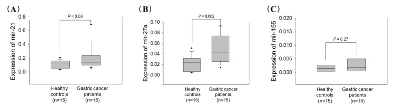
To select potential miRNAs for gastric cancer detection, we divided our samples into the 30 screening cohort and 40 validation cohort. The expression levels of three well-known oncogenic miRNAs were evaluated in 30 plasma samples consisting of 15 patients with gastric cancer and 15 healthy controls. Box plots for the quantitative fluorogenic real-time PCR values are shown in Fig. 1. The miR-21 and miR-155 expression levels were not significantly different between the neoplastic group and the control (p = 0.08 and p = 0.27) (Fig. 1A and 1C). However, the plasma miR-27a expression levels were significantly higher in the patients with gastric cancer (p = 0.002) (Fig. 1B). Although miR-21 expression level in plasma was tend to be increased in patient with gastric cancer compared to healthy control (p = 0.08), we selected miR-27a as a high priority for further analysis.
miR-27a expression level in paired tissue samples
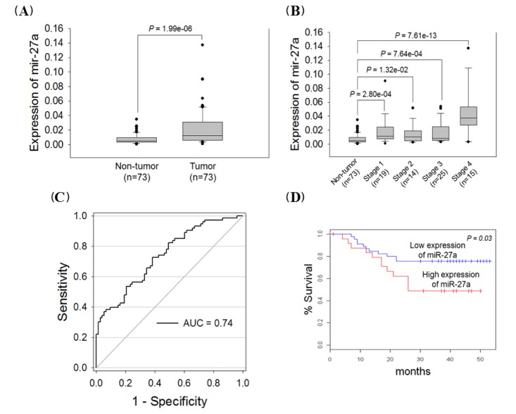
We examined the miR-27a expression level in 73 paired gastric cancer tissues to confirm the oncogenic expression of this miRNA variant. The miR-27a expression level was elevated 2.8-fold in gastric cancer tissues compared with non-tumor tissues (p = 1.96e-06) (Fig. 2A), and this miRNA was highly expressed in 61 out of 73 gastric cancer patient samples (80%). The miR-27a expression levels were highly elevated in all stages compared with non-tumor tissues (Fig. 2B). We divided the patient samples into diffuse and intestinal types, based on the Lauren classification, because gastric cancer patients with diffuse-type tumors have a much poorer prognosis than those with the intestinal type. We observed that the miR-27a expression levels were not significantly different between intestinal and diffuse types (p = 0.19; data not shown). By ROC analysis, we observed that the optimal cut-off value for discriminating between patients and healthy individuals was 0.0067 for a miR-27a expression. miR-27a had 73% sensitivity and 62% specificity in discriminating gastric cancer (Fig. 2C). Importantly, we observed a significant association between miR-27a overexpression and poor survival (p = 0.03) (Fig. 2D), suggesting that miR-27a has potential to be used as a prognostic marker for patients with gastric cancer. Taken together, the miR-27a expression pattern resembled that of an oncogenic microRNA and was associated with gastric cancer patient survival.
MiR-27a expression level in validation cohort
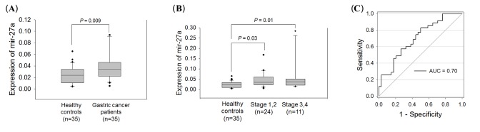
To further validate the diagnostic value of miR-27a, its expression level was measured in additional plasma samples drawn from 20 patients with gastric cancer and 20 healthy controls. The result of miR-27a expression data obtained from each 15 patients with gastric cancer and healthy control plasma samples used in screening cohort were also included in the validation cohort, because of limited sample size. The qRT-PCR results revealed that the miR-27a expression level was highly elevated in the neoplastic group (p = 0.009) (Fig. 3A). In both early (I and II) and late stages (III and IV), the miR-27a expression levels were significantly higher in the group with gastric cancer (Fig. 3B). By ROC analysis, we observed the AUC value to be 0.70 with 75% sensitivity and 56% specificity in discriminating gastric cancer based on the cut-off value of 0.031 for miR-27a expression level in plasma (Fig. 3C). Our results suggest that the plasma miR-27a expression level may have potential as a non-invasive diagnostic biomarker for gastric cancer.
Discussion
A diagnostic source that would provide high specificity and sensitivity may be of enormous benefit to cancer patients, particularly if the biosource could be obtained from non-invasive materials. It was demonstrated that miRNAs are involved in multiple steps of carcinogenesis, either by oncogenic or tumor suppressor functions [31], and circulating miRNAs exist in the serum or plasma [29,32,33]. More importantly, miRNAs have been detected in human serum and plasma in stable forms because of their short hairpin structures, encapsulation by microvesicles, and association with protein complexes [22,23,34,35]. Thus, the detection of any altered expression of tumor-specific miRNAs in saliva, serum, plasma, and urine may offer a promising approach for non-invasive gastric cancer detection.
In this study, the plasma expression levels of three miRNAs (miR-21, miR-27a, and miR-155) were measured in a screening cohort, and we observed that miR-27a expression was significantly elevated in gastric cancer samples compared with healthy control samples. Our result is similar to previously reported results [36]. Next, we investigated whether plasma miR-27a could be released from gastric tumor tissues. We observed that the miR-27a expression level was similar between gastric tumor tissues and plasma, suggesting that the plasma miR-27a expression level may reflect that of the gastric tumor tissues.
miR-27a is located on chromosome 19, and has oncogenic function in gastric adenocarcinoma, by targeting prohibition [16]. This miRNA variant is associated with lymph node metastasis and is overexpressed in gastric cancer [17]. In addition, miR-27a can regulate tumor suppress genes such as SPRY2, and FBW7 [37,38]. SPRY2 can inhibit tumor growth and metastases by interfering with Ras/MAPK activation [37], and FBW7 acts as the substrate recognition component of a Skp1-Cul1-F-box-protein ubiquitin ligase that targets numerous oncoproteins for proteasomal degradation [38]. Growing evidence has suggested that miR-27a has an important role in gastric cancer development and progression.
To estimate its potential value for gastric cancer detection, we evaluated the plasma miR-27a expression level in a validation cohort consisting of 35 patients with gastric cancer and 35 healthy control samples. The sensitivity for the validation cohort was estimated as 75% and the specificity as 56%. Current diagnostic tools for gastric cancer, such as the serological markers carbohydrate antigen 19-9 and carcinoembryonic antigen have low specificity and sensitivity [36]. Our data indicates that the sensitivity and specificity of plasma miR-27a are better than that of the serological markers, although plasma miR-27a alone is not sufficient for accurate gastric cancer detection. Thus, real improvement in the accuracy of gastric cancer detection still needs to be achieved by recruiting additional plasma miRNA markers.
In conclusion, plasma miR-27a could be a useful non-invasive biomarker for gastric cancer detection. Additional studies using a large cohort of samples are needed to validate these results because our study had a small sample size.