Genomics Inform Search

CLOSE


Genomics Inform > Volume 21(1); 2023 > Article
Park, Kim, Kim, and Lee: Generation and analysis of whole-genome sequencing data in human mammary epithelial cells

Abstract

Breast cancer is the most common cancer worldwide, and advanced breast cancer with metastases is incurable mainly with currently available therapies. Therefore, it is essential to understand molecular characteristics during the progression of breast carcinogenesis. Here, we report a dataset of whole genomes from the human mammary epithelial cell system derived from a reduction mammoplasty specimen. This system comprises pre-stasis 184D cells, considered normal, and seven cell lines along cancer progression series that are immortalized or additionally acquired anchorage-independent growth. Our analysis of the whole-genome sequencing (WGS) data indicates that those seven cancer progression series cells have somatic mutations whose number ranges from 8,393 to 39,564 (with an average of 30,591) compared to 184D cells. These WGS data and our mutation analysis will provide helpful information to identify driver mutations and elucidate molecular mechanisms for breast carcinogenesis.

Introduction

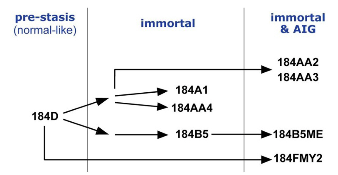
Breast cancer is the most common cancer diagnosed among women in the United States (excluding skin cancers). It is the second leading cause of cancer death among women after lung cancer [1]. It is curable in ~70%–80% of early-stage patients before metastasis. However, advanced breast cancer with distant organ metastases is considered incurable with currently available therapies [2]. Therefore, it is crucial to understand molecular characteristics that are associated with the development of breast cancer and to identify molecular biomarkers. A cell-based model system is essential for an in-depth study of molecular events during the human breast tumorigenesis. Human mammary epithelial cell (HMEC) lines, developed from normal breast tissues, are an ideal in vitro cell line model recapitulating early events of breast tumorigenesis [3] (see also https://hmec.lbl.gov/mock/history.html). Briefly, 184D is primary culture cells obtained from the reduction mammoplasty specimen 184. Most 184D cells underwent cell death (so-called the stasis barrier). 184D cells were treated with a mutagen benzo[a]pyrene or transformed by c-MYC transduction to overcome the stasis barrier. They were clonally selected to yield seven HMEC lines ([4-8], summarized in Fig. 1). In this study, we obtained and analyzed whole-genome sequencing (WGS) data from HMEC lines, which will help understand early breast carcinogenesis at the genomic level.

Methods

Cell line and other reagents

HMEC cultures were derived and grown as previously published [5-7,9]. The sources of other reagents were described in our previous study [10].

WGS library construction and sequencing

We used the QIAamp DNA Mini Kit (Qiagen, Carlsbad, CA, USA) to isolate gDNA from HMEC cultures. The quantity of the extracted gDNA was analyzed with an ND-1000 spectrophotometer (Thermo Fisher Scientific, Waltham, MA, USA). For WGS library construction, we used the TruSeq DNA library Prep Kit (Illumina, San Diego, CA, USA) according to the manufacturer’s instructions. For WGS, paired-end sequencing was performed on the Illumina HiSeq X Ten sequencing instrument, yielding ~150-bp short sequencing reads.

Data analysis

Raw sequence reads were aligned to the human reference genome 19 using Burrows Wheelers Aligner [11], and duplicate reads were removed using Picard (Broad Institute). We used Qualimap 2 to evaluate next-generation sequencing alignment data [12]. Then, the remaining reads were calibrated and realigned using the Genome Analysis Toolkit [13]. The realigned Binary Alignment Map files were analyzed using Strelka2 [14] to detect somatic single-nucleotide variants and insertions/deletions. The relative distribution of single-base substitutions was analyzed by the Maftools [15]. We used HOMER to annotate somatic mutation to the hg19 genome [16]. For driver mutation analysis, we download the driver gene list from the IntOgen cancer mutation browser [17]. For all programs, we used the default parameter setting.

Data availability

The whole-genome data are available in the Korean Nucleotide Archive (KoNA, https://kobic.re.kr/kona) and Sequence Read Archive (SRA, https://www.ncbi.nlm.nih.gov/sra) public database with the accession number PRJKA220370 and PRJNA913438.

Results and Discussion

Quality and quantity of the sequencing data

We performed WGS on a total of eight HMEC cultures (shown in Fig. 1): pre-stasis 184D, its derivatives immortalized cell lines (184A1, 184AA4, and 184B5), and immortalized ones that further acquired AIG (184AA2, 184AA3, 184B5ME and 184FMY2). First, we assessed the quality and quantity of the WGS data, including mapping rates, genome coverage, scores of the mapping quality, and duplicate reads using Qualimap 2. These values are summarized in Table 1. Briefly, the mapping rate and scores of the mapping quality of the eight samples were higher than 85% and 53%, respectively. In addition, the average genome coverage was more than 30× (between 31.84× and 42.84×) in all eight samples. WGS data with 30× sequence coverage is appropriate for comprehensively identifying tumor-specific somatic mutations [18]. These results indicate that the quality and quantity of our WGS data were satisfactory for mutational analysis in HMEC cultures.

Mutation patterns identified from the HMEC model

We analyzed somatic mutations from the WGS data. 184D cells are the primary culture of normal breast tissue and yet-to-be immortalized. Therefore, we considered 184D as normal breast tissue and used its genome sequence as a reference sequence when analyzing WGS data of the other seven HMEC lines that are cancer progression series.
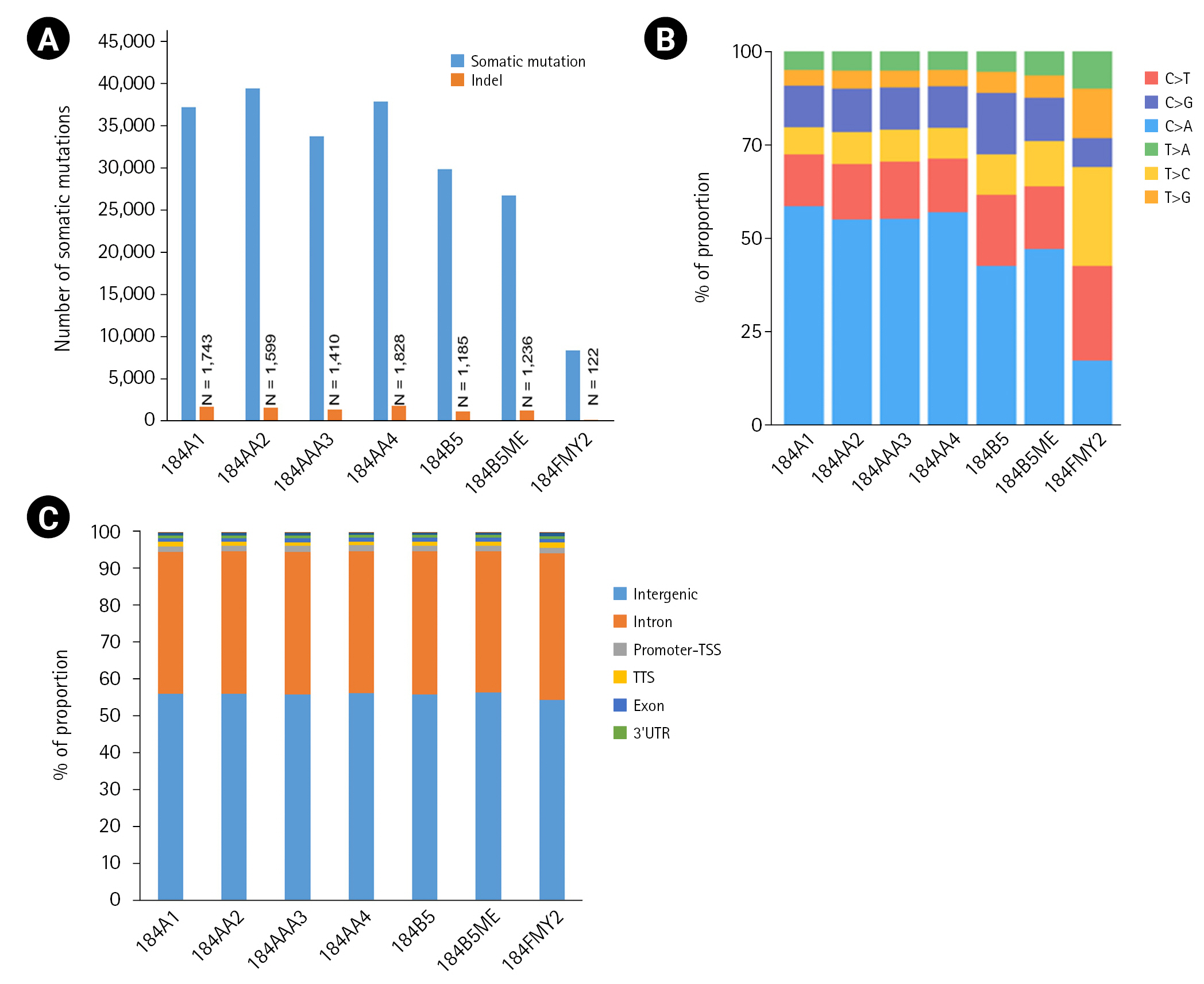
Among the seven HMEC lines, the number of somatic mutations per sample ranged from 8,393 to 39,564, with an average of 30,591 (Fig. 2A). In particular, 184FMY2 had notably low somatic mutation frequency (n = 8,393), in agreement with the fact that it had been made by c-MYC transduction, whereas the other HMEC lines were treated with benzo[a]pyrene. Next, we examined the pattern of base substitutions. Except for 184FMY2, we observed that ~50% of mutations were C>A and that ~30% were C>T and C>G transversions (Fig. 2B), similarly to a previous study [19]. We annotated those somatic mutations to the hg19 reference genome and observed that most somatic mutations were located in the intergenic and intronic regions (Fig. 2C).
Since non-synonymous mutations are likely to be essential and are functionally annotatable, we focused on them. The number of mutations affecting protein-coding genes was 52 to 361 in each sample (data not shown). Then, we performed the driver mutation analysis using the IntOGen cancer mutation browser [17] and observed that 36 non-synonymous mutations in the HMEC cancer progression series coincided with the cancer driver mutations (Table 2). Further study will be needed to validate whether the mutated genes are genuinely associated with breast carcinogenesis.
In this study, we generated WGS data and analyzed mutation profiles in the HMEC cancer progression series because genetic mutations are one of the most significant factors in determining breast cancer progression and therapeutic management [20]. We hope that our WGS data of HMEC lines will provide useful information to breast cancer researchers and clinicians.

Notes

Authors’ Contribution

Conceptualization: SYK, YSL.

Sample and data curation: JLP, SYK, YSL.

WGS data generation and analysis: JLP, JYK.

Writing - original draft: JLP.

Writing - review & editing: SYK, YSL.

Conflicts of Interest

No potential conflict of interest relevant to this article was reported.

Acknowledgments

This work was supported by grants from: National Research Foundation of Korea (NRF) funded by the Korea government (MEST) (NRF-2017M3A9B5060884 to J-LP and S-YK) and the National Cancer Center Korea (NCC-2110190 to YSL).

Fig. 1.
Diagram illustrating the human mammary epithelial cell progression series derived from a reduction mammoplasty specimen 184.
gi-22044f1.jpg
Fig. 2.
The number of somatic mutations and distribution of mutation types. (A) Somatic mutations were detected using the Strelka2 package with the default parameter setting. (B) Relative distribution of single-base substitutions by type in each human mammary epithelial cell culture sample. (C) Distribution of somatic mutation in the genome. Somatic mutations were annotated to the hg19 using the HOMER package. UTR, untranslated region.
gi-22044f2.jpg
Table 1.
Quality and quantity of the sequencing data
Sample ID Total No. of reads Mapped reads, n (%) Duplicate reads, n (%) Genome coverage (mean) Mapping quality
184A1 913,043,618 853,249,715 (93.45) 118,589,417 (12.99) 40.62 54.03
184AA2 870,033,090 826,770,914 (95.03) 101,833,624 (11.7) 39.31 53.90
184AA3 819,634,530 733,795,759 (89.53) 95,136,431 (11.61) 34.95 53.98
184AA4 794,654,212 762,287,813 (95.93) 102,740,630 (12.93) 36.31 53.98
184B5 1,008,170,478 898,909,936 (89.16) 164,770,658 (16.34) 42.84 53.96
184B5ME 799,810,062 734,277,857 (91.81) 92,112,929 (11.52) 35.00 54.08
184D 779,003,944 669,465,976 (85.94) 86,326,513 (11.08) 31.87 54.00
184FMY2 828,214,280 791,321,104 (95.55) 117,711,972 (14.21) 37.69 54.02
Table 2.
Annotation of non-synonymous mutations in each of HMEC samples to a cancer driver mutations database
Symbol Immortal
Immortal with AIG
184A1 184AA4 184B5 184AA2 184AA3 185B5ME 184FMY2
MTOR Q1627K Q1627K ND Q1627K Q1627K ND ND
CSF3R T154N T154N ND T154N T154N ND ND
KMT2D M1417I M1417I ND M1417I M1417I ND ND
ACSL3 G476V G476V ND G476V G476V ND ND
PLCG1 D342Y D342Y ND D342Y D342Y ND ND
CARD11 T43M T43M ND T43M T43M ND ND
AMER1 P49Q, P49T P49Q, P49T ND P49Q, P49T P49Q, P49T ND ND
BTK R332S R332S ND R332S R332S ND ND
NFKBIE K316N ND ND K316N K316N ND ND
SETBP1 D412H D412H ND D412H ND ND ND
CDKN2A G102V G102V ND G102V ND ND ND
MED12 M880I M880I ND ND M880I ND ND
RSPH10B2 S71T ND ND ND S71T S71T ND
FOXD4L1 ND ND P234R Y110S, P234R ND Y110S ND
CIITA ND ND Q444K ND ND Q444K ND
ZNF626 ND ND G253R ND ND G253R ND
CUL3 ND ND G283V ND ND G283V ND
MYH9 ND ND L171F ND ND L171F ND
FGD5 ND ND G395C ND ND G395C ND
NPRL2 ND ND A141S ND ND A141S ND
TET2 ND ND E1144V ND ND E1144V ND
ABCB1 ND ND R905G ND ND R905G ND
CDH11 ND R218I ND R28Q ND ND ND
PDPR ND ND ND ND ND I47V I47V
H3F3A A115G ND ND ND ND ND ND
CIC T1560P ND ND ND ND ND ND
NOTCH2 ND ND P210L ND ND ND ND
PEG3 ND Q1182H ND ND ND ND ND
KDM3B ND ND N1092Y ND ND ND ND
LRP1B ND ND G838R ND ND ND ND
PML ND ND L825V ND ND ND ND
CLTCL1 ND ND E1304Q ND ND ND ND
RHPN2 ND ND ND ND K216R ND ND
FANCD2 ND ND ND ND P87R ND ND
ZNF429 ND ND ND ND ND ND S498R

HMEC, human mammary epithelial cell; ND, not detected.

References

1. DeSantis CE, Ma J, Gaudet MM, Newman LA, Miller KD, Goding Sauer A, et al. Breast cancer statistics, 2019. CA Cancer J Clin 2019;69:438–451.
crossref pmid pdf
2. Harbeck N, Penault-Llorca F, Cortes J, Gnant M, Houssami N, Poortmans P, et al. Breast cancer. Nat Rev Dis Primers 2019;5:66.
crossref pmid pdf
3. Labarge MA, Garbe JC, Stampfer MR. Processing of human reduction mammoplasty and mastectomy tissues for cell culture. J Vis Exp 2013;(71):50011.
crossref
4. Hines WC, Kuhn I, Thi K, Chu B, Stanford-Moore G, Sampayo R, et al. 184AA3: a xenograft model of ER+ breast adenocarcinoma. Breast Cancer Res Treat 2016;155:37–52.
crossref pmid pdf
5. Lee JK, Garbe JC, Vrba L, Miyano M, Futscher BW, Stampfer MR, et al. Age and the means of bypassing stasis influence the intrinsic subtype of immortalized human mammary epithelial cells. Front Cell Dev Biol 2015;3:13.
crossref pmid pmc
6. Garbe JC, Vrba L, Sputova K, Fuchs L, Novak P, Brothman AR, et al. Immortalization of normal human mammary epithelial cells in two steps by direct targeting of senescence barriers does not require gross genomic alterations. Cell Cycle 2014;13:3423–3435.
crossref pmid pmc
7. Stampfer MR, Garbe J, Nijjar T, Wigington D, Swisshelm K, Yaswen P. Loss of p53 function accelerates acquisition of telomerase activity in indefinite lifespan human mammary epithelial cell lines. Oncogene 2003;22:5238–5251.
crossref pmid pdf
8. Stampfer MR, Bartley JC. Induction of transformation and continuous cell lines from normal human mammary epithelial cells after exposure to benzo[a]pyrene. Proc Natl Acad Sci U S A 1985;82:2394–2398.
crossref pmid pmc
9. Garbe JC, Bhattacharya S, Merchant B, Bassett E, Swisshelm K, Feiler HS, et al. Molecular distinctions between stasis and telomere attrition senescence barriers shown by long-term culture of normal human mammary epithelial cells. Cancer Res 2009;69:7557–7568.
crossref pmid pmc pdf
10. Park JL, Lee YS, Song MJ, Hong SH, Ahn JH, Seo EH, et al. Epigenetic regulation of RNA polymerase III transcription in early breast tumorigenesis. Oncogene 2017;36:6793–6804.
crossref pmid pdf
11. Li H, Durbin R. Fast and accurate long-read alignment with Burrows-Wheeler transform. Bioinformatics 2010;26:589–595.
crossref pmid pmc pdf
12. Garcia-Alcalde F, Okonechnikov K, Carbonell J, Cruz LM, Gotz S, Tarazona S, et al. Qualimap: evaluating next-generation sequencing alignment data. Bioinformatics 2012;28:2678–2679.
crossref pmid pdf
13. McKenna A, Hanna M, Banks E, Sivachenko A, Cibulskis K, Kernytsky A, et al. The Genome Analysis Toolkit: a MapReduce framework for analyzing next-generation DNA sequencing data. Genome Res 2010;20:1297–1303.
crossref pmid pmc
14. Kim S, Scheffler K, Halpern AL, Bekritsky MA, Noh E, Kallberg M, et al. Strelka2: fast and accurate calling of germline and somatic variants. Nat Methods 2018;15:591–594.
crossref pmid pdf
15. Mayakonda A, Lin DC, Assenov Y, Plass C, Koeffler HP. Maftools: efficient and comprehensive analysis of somatic variants in cancer. Genome Res 2018;28:1747–1756.
crossref pmid pmc
16. Heinz S, Benner C, Spann N, Bertolino E, Lin YC, Laslo P, et al. Simple combinations of lineage-determining transcription factors prime cis-regulatory elements required for macrophage and B cell identities. Mol Cell 2010;38:576–589.
crossref pmid pmc
17. Gonzalez-Perez A, Perez-Llamas C, Deu-Pons J, Tamborero D, Schroeder MP, Jene-Sanz A, et al. IntOGen-mutations identifies cancer drivers across tumor types. Nat Methods 2013;10:1081–1082.
crossref pmid pmc pdf
18. Alioto TS, Buchhalter I, Derdak S, Hutter B, Eldridge MD, Hovig E, et al. A comprehensive assessment of somatic mutation detection in cancer using whole-genome sequencing. Nat Commun 2015;6:10001.
crossref pmid pdf
19. Severson PL, Vrba L, Stampfer MR, Futscher BW. Exome-wide mutation profile in benzo[a]pyrene-derived post-stasis and immortal human mammary epithelial cells. Mutat Res Genet Toxicol Environ Mutagen 2014;775-776:48–54.
crossref pmid
20. Shiovitz S, Korde LA. Genetics of breast cancer: a topic in evolution. Ann Oncol 2015;26:1291–1299.
crossref pmid pmc


ABOUT
ARTICLE CATEGORY

Browse all articles >

BROWSE ARTICLES
FOR CONTRIBUTORS
Editorial Office
Room No. 806, 193 Mallijae-ro, Jung-gu, Seoul 04501, Korea
Tel: +82-2-558-9394    Fax: +82-2-558-9434    E-mail: kogo3@kogo.or.kr                

Copyright © 2024 by Korea Genome Organization.

Developed in M2PI

Close layer
prev next